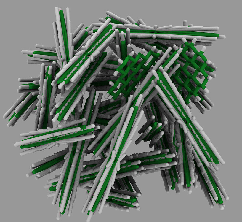

7 In addition, numerous compounds have been found to reduce Aβ 1−42 aggregation in vitro. Some reports have demonstrated that the presence of Cu 2+ favors fibril formation, 10 while other recent studies suggested that Cu 2+ might inhibit Aβ 1−42 fibril formation.

However, although some research has shown that Aβ contains amino acid residues that can interact with Cu 2+, 9 the effects of these interactions with respect to the Aβ 1−42 oligomerization process have been contradictory. 8 Therefore, several experimental and computational methods have focused on the analysis of the interaction between Aβ 1−42 and Cu 2+. 6, 7 The interaction between Aβ-fibrils and Cu 2+ is important not only because of its role in amyloidosis but also because it has been associated with the generation of oxidative stress. One of the metals that has attracted attention is copper (Cu 2+), whose concentration is threefold greater in AD patients than in normal patients, with concentrations between 340 and 400 µ M. Several studies have explored the use of metals and small compounds as oligomerization inhibitors, but a molecular-level understanding of the inhibition mechanism remains unclear. 5īecause of the toxicity of Aβ 1–42 oligomers and fibrils, the prevention of amyloidosis could have an enormous impact on the treatment of AD. Furthermore, during these processes, the β-sheet conformation is defined and stabilized by the formation of an intrachain and interchain salt bridge between Asp 23/Glu 22 and Lys 28, increasing the ability of Aβ to nucleate and form fibrils. Aβ 1−42 is composed primarily of hydrophobic amino acid residues at the C-terminal end, 4 which establish hydrophobic interactions during the oligomerization and fibrillization process. 3 The length of the Aβ peptide released by β-secretase varies from 39 to 43 amino acid residues, with the 42 amino acid residue peptide (Aβ 1−42) being the most commonly found in fibrils. Aβ is produced from the proteolytic cleavage of amyloid precursor protein (APP) by β and γ secretases or by α and γ-secretases to initiate amyloidogenic or nonamyloidogenic pathways, respectively. 2 Amyloid fibrils and oligomers are formed by a process called amyloidosis, in which the beta-amyloid peptide (Aβ) produces insoluble aggregates.


The accumulation of insoluble amyloid “plaques” and oligomers of beta amyloid peptide (Aβ) in the brain plays an important role in Alzheimer's disease (AD), which is one of the principal causes of dementia worldwide, 1 causing oxidative stress and inflammation in specific areas of the brain. Then, ligands that bind Asp 23 or Glu 22 and Lys 28 could therefore be used to prevent β turn formation and, consequently, the formation of Aβ fibrils. The in vitro studies demonstrated that Aβ remains in an unfolded conformation when Cu 2+ and galanthamine are used. The docking results revealed that the conformation obtained by the MD simulation of a monomer from the 1Z0Q structure can form similar interactions to those obtained from the 2BGE structure in the oligomers. However, the MD simulations of Aβ 1−42 in the presence of Cu 2+ or galanthamine demonstrated that both ligands prevent the formation of the salt bridge by either binding to Glu 22 and Asp 23 (Cu 2+) or to Lys 28 (galanthamine), which prevents Aβ 1−42 from adopting the U-characteristic conformation that allows the amino acids to transition to a β-sheet conformation. The formation of a salt bridge between Asp 23 and Lys 28 was also observed beginning at 5 ns. The MD simulations revealed that Aβ 1–42 acquires a characteristic U-shape before the α-helix to β-sheet conformational change. Therefore, the aim of this work was to explore how Cu 2+ and galanthamine prevent the formation of Aβ 1–42 fibrils using molecular dynamics (MD) simulations (20 ns) and in vitro studies using fluorescence and circular dichroism (CD) spectroscopies. However, the mechanism of this inhibition remains unclear. Recently, Cu 2+ and various drugs used for AD treatment, such as galanthamine (Reminyl®), have been reported to inhibit the formation of Aβ fibrils. The formation of Aβ fibrils and oligomers requires a conformational change from an α-helix to a β-sheet conformation, which is encouraged by the formation of a salt bridge between Asp 23 or Glu 22 and Lys 28. The formation of fibrils and oligomers of amyloid beta (Aβ) with 42 amino acid residues (Aβ 1–42) is the most important pathophysiological event associated with Alzheimer's disease (AD).


 0 kommentar(er)
